Our Research
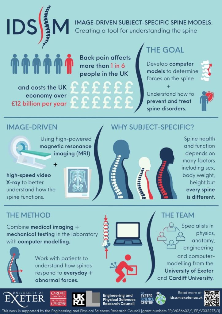
Find out more about the project motivation and aims in our latest infographic, and learn more about the research methods and facilities below.
Background
Research Methods
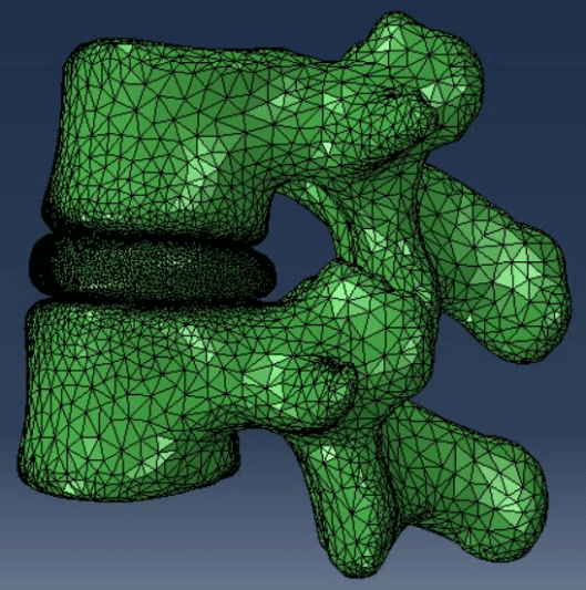
This multi-disciplinary project involves imaging spines and using this information to design a computer model that can be customised for each patient.
The imaging needs to be carried out in various ways, to get a complete picture of how the spine moves and responds to stresses. The different members of the team will be testing spine samples mechanically (in vitro testing), imaging volunteers (in vivo) and measuring how their spines move during different activities, and imaging samples and volunteers using magnetic resonance imaging (MRI). The computer model will be developed based on this imaging data.
Find out more about the research methods below.
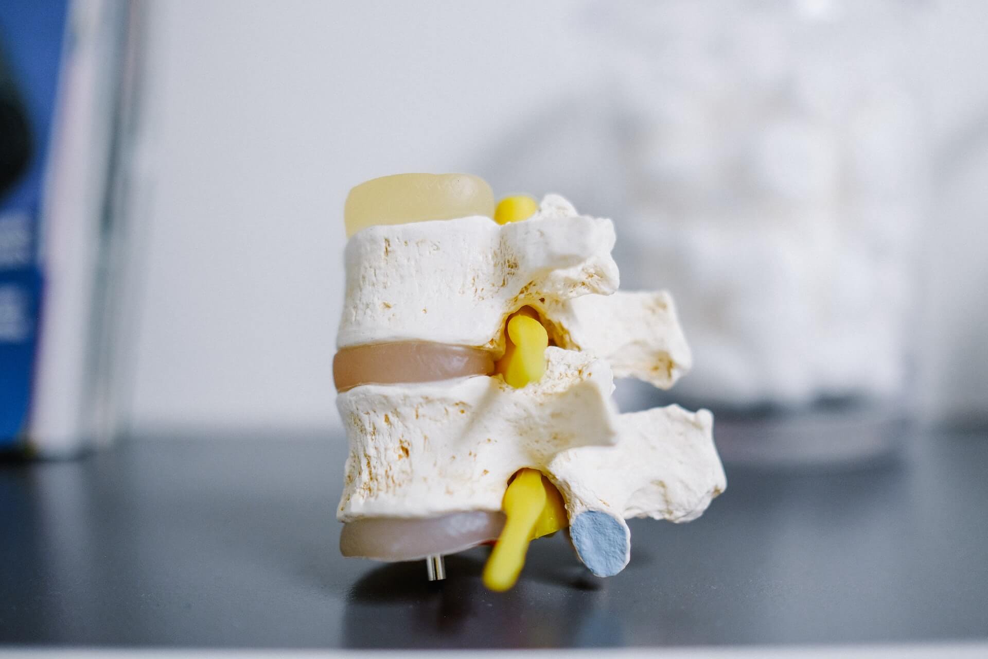
In vitro testing
This part of the project investigates the loads (forces) on the spine using a six-axis spine simulator,
In the first part of the project, the team will apply forces to the samples and use pressure sensors to understand how the motion corresponds to pressure in the discs and joints. This information will be combined with the MRI data and used to inform the computer model. In later stages of the project, the team can apply forces to the spinal samples to replicate the loads that people encounter in everyday activities, which can then be fed back into the computer model.
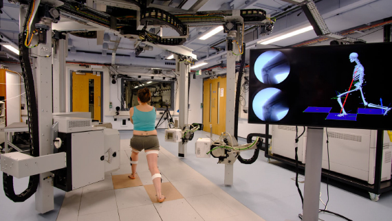
In vivo testing (using bi-plane fluoroscopy)
Bi-plane fluoroscopy is a new technique sometimes called video radiography. The technique uses two X ray machines on either side of the patient taking a quick series of short X rays – described as ‘pulsed’ X rays. In this study, the participants will perform a series of movements similar to those that happen during day to day living, e.g. bending forwards or picking up objects. As the participant is moving, the X rays capture precisely how the internal structures of the body respond and react to the movement.
The X ray images are taken at up to 125 frames per second, providing a highly accurate mini movie or video of the movements. This helps us to build the 3D model of exactly what is happening to the spine and the surrounding tissue.
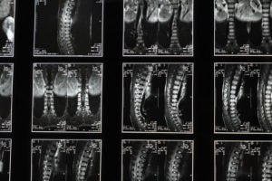
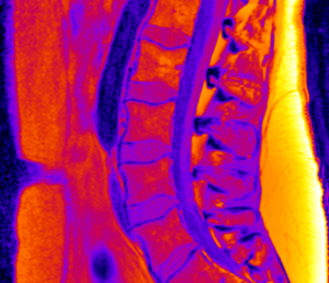
Magnetic Resonance Imaging (MRI)
This imaging technique is a useful way to probe internal structures, both in vitro (samples) and in vivo (patients). It uses a magnetic field that changes in time to give a short burst of energy to the hydrogen atoms in the sample / person (which are mostly in water and fat). When the hydrogen atoms give out this energy again this is measured by the MRI machine, and using software, this can be made into an image. By comparing the different parts of the image, scientists can identify the parts of the sample which have different concentrations of water / fat – which correspond to different materials (like tissue, ligaments, bone, etc.)
As well as getting a 3D image of the spine and its surroundings, an important part of this study is to develop the method for estimating the material properties from the MRI data.
Computer modelling
A computer model is an approximate, digital version of a real thing – in this case, the spine and surrounding tissue. Unlike a piece of software that has already been programmed to be able to model a building or a piece of machinery for example, the spinal model is completely new, and has to be constructed based on real-world data – from the in vitro, in vivo and MRI studies.
The 3D image and material properties from the MRI scan can be used to build the physical properties of the model. The especially challenging part is programming the model so that the bones, discs, ligaments or muscle can respond to motion or stresses accurately. The team will make use of machine learning to analyse the data and work out how to make the model as accurate as possible.
Facilities
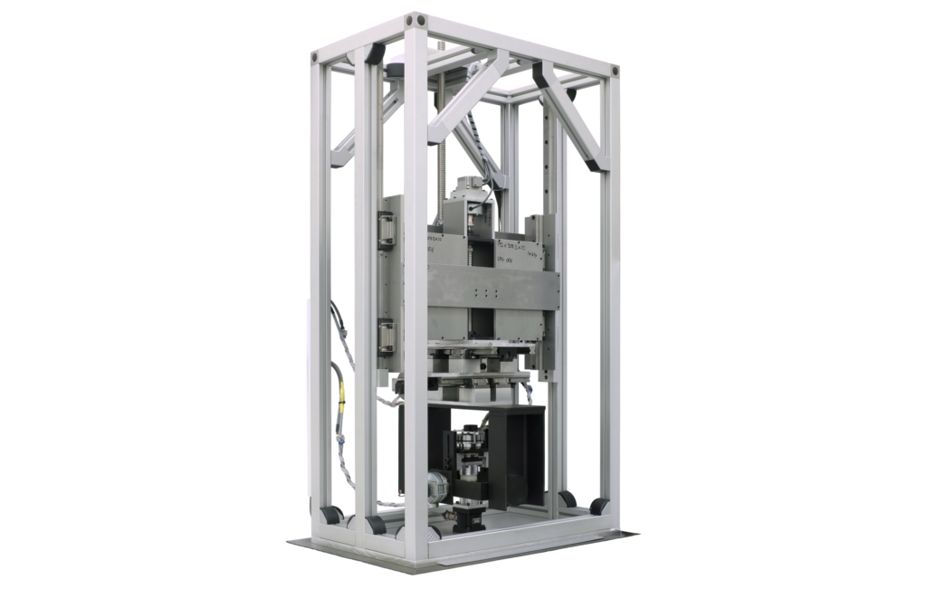
University of Exeter Biomechanical Engineering Research Laboratory: Six-Axis Spine Simulator
The six-axis spine simulator is a new piece of equipment being developed, which can apply translation and rotation forces to spinal samples in 6 different ways. It comprises a hydraulic testing machine and a translation platform, so can apply extension, compression, rotations and bending forces to samples.
Cardiff University Musculoskeletal Biomechanics Research Facility: Bi Plane X Ray Imaging
This is a world-leading facility for studying biomechanics and musculoskeletal health. The research team will be using the facility’s fluoroscopy laboratory for in vivo testing, described above. This lab is unique in the UK, and one of only a handful of similar facilities worldwide.
To find out more, check out the facility’s webpage here.